The 2026 Nobel Prize in Medicine, explained

A light switch for the brain

Your brain is built from about 86 billion tiny living wires called nerve cells, of many different kinds, packed together like tangled spaghetti. For a hundred years nobody could switch just one kind on or off, so nobody could find out which cells make you feel scared, sleepy or thirsty.

In one line

Three scientists found a light-opened gate, a tiny door in a cell’s skin, in a pond alga and fitted it into chosen brain cells, so a flash of light can now switch exactly those cells on or off.

Short on time? Read the 5 key things or jump to the game. New to how the brain works? Start with Brain Basics.

Start here

The whole story in six pictures

If you only look at one thing on this page, look at this.
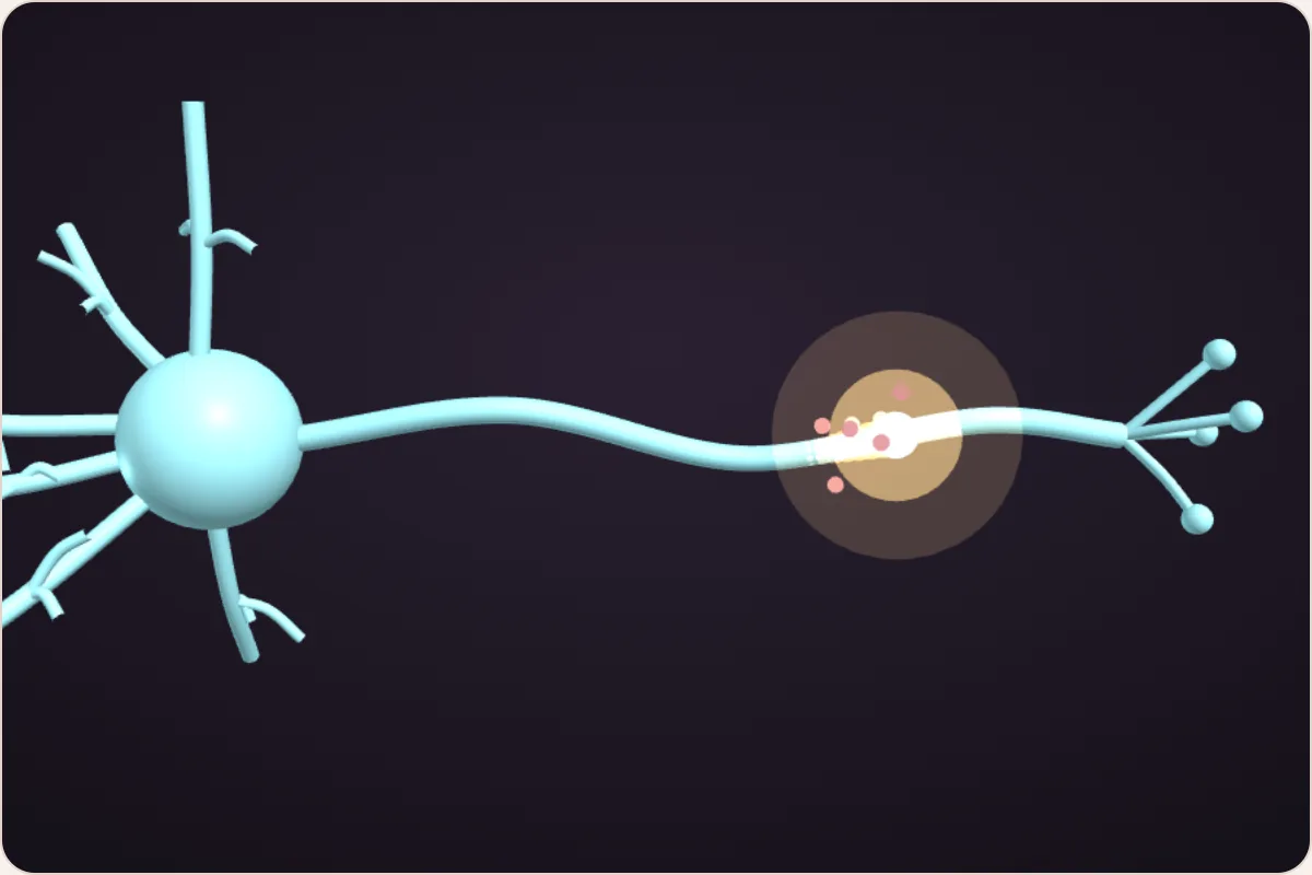
A 3D nerve cell firing: a bright electric spark races along its long wire, the axon 1 · Tiny sparks

Brain cells talk with tiny electric sparks. A spark starts when little gates in the cell’s skin open.
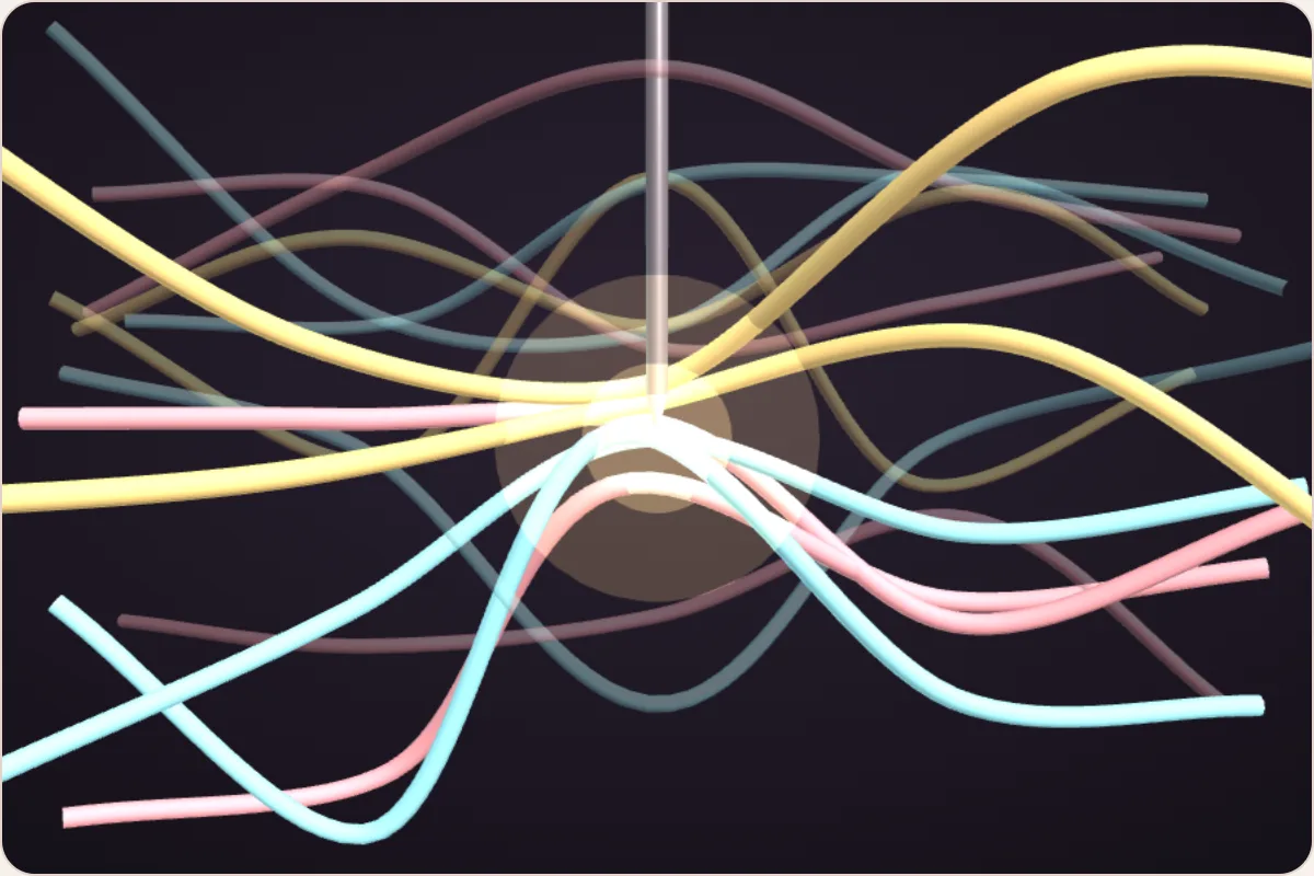
A 3D tangle of nerve-cell wires in three colours, packed together, with a metal electrode in the middle lighting up every wire near its tip 2 · A tangle

Many kinds of brain cell sit tangled together. The old tools switched on all of them at once.
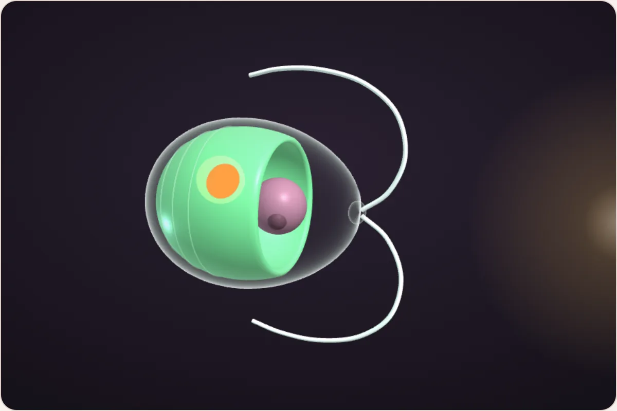
A 3D model of the one-celled alga Chlamydomonas: a see-through cell with a green cup inside, an orange eyespot and two long tails 3 · The alga

A one-celled pond alga swims toward light, and reacts to it astonishingly fast.
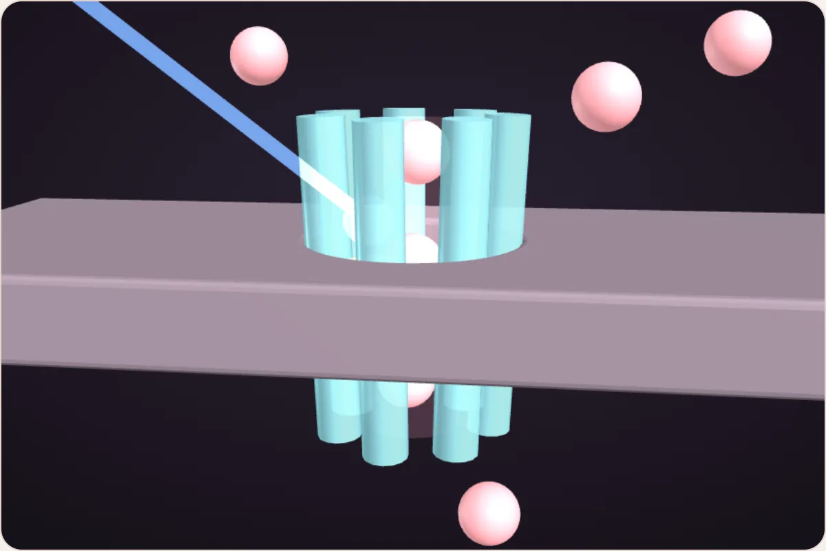
3D channelrhodopsin: a beam of blue light hits a teal gate set in the cell’s skin, and pink sodium particles pass through it into the cell 4 · The gate, 2002–2003

Its light catcher is itself a gate: light hits, it opens. Frog eggs given its gene (its recipe) proved it.
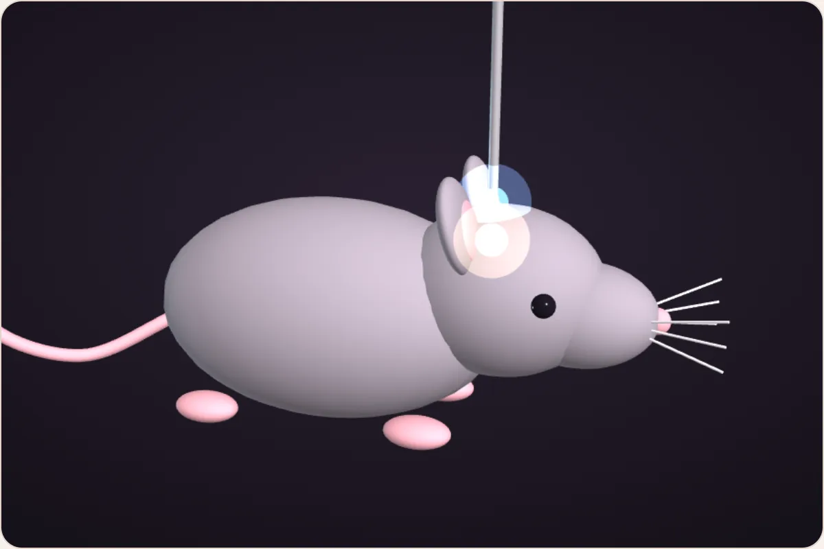
A 3D mouse with a hair-thin glass fibre glowing blue into its head, where a small group of cells lights up 5 · The switch, 2005–2007

Put the alga’s recipe into one kind of brain cell, and a flash of blue light switches just those cells on.
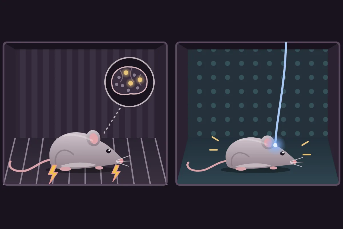
The fear-memory experiment: a mouse gets a mild shock in a striped box; later, in a safe spotted box, blue light on a few brain cells makes it freeze 6 · What it unlocked

A remote control for chosen cells: light on a few cells brought back a mouse’s memory of fear, and in a blind man’s eye, light gates stand in for lost cells to give back some sight.

Chapter 1

Your brain runs on tiny sparks

The full explanation, from zero: Brain Basics: nerve cells · the cell’s charge · gates · how a nerve cell fires

To understand this prize you need just one idea about how brain cells work. Here it is in three steps.

  1. Step 1Nerve cellsYour brain is built from about 86 billion nerve cells: cells shaped like wires, with short branches that collect messages and one long wire, the axon, that sends a message on.
  2. Step 2Each cell is chargedYour body’s water is full of charged particles (bits of dissolved salt, also called ions). Some carry a plus charge, some a minus charge. A nerve cell keeps them unevenly spread across its skin, the membrane, so its inside is slightly negative, like a small battery.
  3. Step 3Gates start the sparkThe skin has tiny tunnels called gates (scientists say ion channels). When enough open, plus particles rush in and flip the inside from negative to positive for a moment. That quick flip is a spike, a tiny electric spark lasting about a thousandth of a second, which races down the axon and passes the message on.

So switching a nerve cell on means opening its gates. In the brain, most gates open for messages arriving from neighbouring cells. Nobody could reach in and open the gates of one particular kind of cell, on purpose, whenever they liked.

Keep that in mind. The whole prize is about a new way to open those gates: with a flash of light.

outside the cell gate shutgate open: spark inside the cell · pink = plus particles
Key idea

Brain cells talk with tiny electric sparks. A spark starts when little gates in the cell’s skin open and let charged particles rush in.

Quick check

What starts a nerve cell’s spark?

Chapter 2

The switch scientists wished for

If you could open the gates of one kind of nerve cell yourself, you could switch those cells on and watch what happens. For most of the last century, nobody could.

Inside your brain, many kinds of nerve cell sit packed together, tangled like spaghetti. A cell that helps make you thirsty can sit right next to one that helps wake you up.

Scientists could watch them. Brain scans and thin metal wires called electrodes show which cells are busy when something happens. But being busy at the same time doesn’t prove a cell causes it. Umbrellas go up whenever it rains, but opening an umbrella doesn’t make it rain.

Scientists could also tell many kinds of nerve cell apart, because each kind reads its own set of genes and makes its own chemicals (Brain Basics: brain wiring). So they often had suspects: a kind of cell that was busy at the right moments, or whose loss caused a problem. What they couldn’t do was test a suspect on its own.

The clearest test is to switch one kind of cell on, by itself, and see what changes. If a calm mouse acts scared the moment you switch on certain cells, those cells can cause fear.

umbrellas up rain together, but not the cause

The tools scientists had for switching cells on were blunt. A metal wire zaps every cell near its tip, whatever its kind. A drug travels through the blood and reaches every cell that has the right receptor (a lock that the drug’s tiny particles fit), and it takes minutes to work. Try both below on a patch of a mouse’s brain. A small group of gold cells there moves the mouse’s whiskers: zap with each tool and watch what else happens.

teal = ear cells rose = leg cells gold = whisker cells (they move the whiskers)

Three kinds of nerve cell, tangled together. The gold ones move the mouse’s whiskers. Pick a tool and try to make only the whiskers twitch.

In 1979 Francis Crick, one of the scientists who discovered the shape of DNA, wrote that brain research badly needed a way to control one kind of cell and leave all the others alone. Twenty years later he guessed that light might be ideal: nerve signals are extremely fast, and light can be flicked on and off just as fast and aimed at one spot.

The problem: brain cells don’t react to light at all, and nobody knew how to make them. Crick himself admitted the idea sounded far-fetched, though perhaps not impossible.

“switch on just one kind”
Curious? Go deeperWhy would light make such a good switch?

Nerve signals are fast: a spike is over in about a thousandth of a second. Light can be switched on and off just as fast, so it could start a cell firing at exactly the right moment, even one spike at a time. It can be aimed at one small spot. And because brain cells normally ignore light, shining it would not disturb anything else, as long as only the chosen cells could react to it.

Key idea

To learn what one kind of brain cell does, you have to switch on just that kind, quickly. The old tools switched on everything nearby.

Quick check

Why wasn’t a metal wire good enough for testing what one kind of cell does?

Chapter 3

A pond alga that swims toward light

The answer to Crick’s wish didn’t come from brain research. It came from a scientist studying pond slime: a living thing that reacts to light, which brain cells can’t do.

Chlamydomonas (say “klam-ee-DOM-oh-nas”) is a tiny green alga that lives in ponds and wet soil. Each one is a single cell, about a hundredth of a millimetre across. It swims with two little tails, and it has an orange spot, the eyespot, that catches light. In the 3D model, which you can drag to turn, the orange dot is the eyespot and the big green cup is the part that makes food from sunlight.

Shine a light at one side of a dish of them and the whole green cloud drifts toward it. An alga lives on sunlight, so swimming toward light keeps it fed. Drag the lamp around the dish below and watch them follow.

eyespottwo tailsone whole living cell
one algaeyespotlamp light

Try it: drag the lamp around the dish, or tap the rim. Each alga’s orange eyespot catches the light, and it steers its two tails toward it, so the whole green cloud gathers on the bright side.

How does a single cell “see”? Every cell is full of tiny machines it builds for itself, called proteins (Brain Basics: proteins). The gates in a nerve cell are proteins. So are the light catchers in your eyes, and in the alga’s eyespot. A light catcher holds a small molecule (a tiny cluster of atoms) called retinal, made from vitamin A, which changes shape when light hits it. In the animation, the gold chain is retinal, sitting inside a lilac light-catching protein: watch it snap into a new shape when light arrives. Each light catcher is tuned to certain colours: the protein wrapped around the retinal decides which colour of light makes it snap most easily. That is how the three kinds of colour-sensing cells in your eyes tell colours apart (Brain Basics: light and colour). The alga’s light catcher reacts most to blue light.

In the early 1990s Peter Hegemann, working at a research institute in Germany, measured the tiny electric signal the alga makes when light reaches its eyespot. It came astonishingly fast: in about half a thousandth of a second.

Your eyes are much slower. In your eye, the light catcher only starts a relay: it nudges a second protein, which nudges a third, and so on like a row of dominoes, and only at the end of the chain do gates change. That takes about a hundredth of a second or more, at least 20 times longer. Press “Flash light” below to race them.

light catcher (a protein)retinal
lightprotein switched oncharged particles
slowed down about 400×

Try it: press “Flash light” to hit both cells with the same flash at the same moment, and watch which one gets its signal out first.

So few steps fit into half a thousandth of a second that Hegemann suspected there was only one: a single protein that catches the light and is itself the gate. Light hits, the gate opens, charged particles flow, done. Nobody had ever seen a gate that light could open on its own, and many scientists doubted it.

The animation shows his idea. The teal barrel is one protein set in the cell’s skin, with retinal (gold) inside it. Blue light snaps the retinal into a bend, the barrel opens into a tunnel, and sodium (pink) pours into the cell.

one protein: light catcher and gate
Curious? Go deeperIs the alga’s light catcher the same as the one in our eyes?

They are distant cousins. Both are proteins holding the same light-catching molecule, retinal, and in both, light makes retinal snap into a new shape. In your eyes, that snap only starts a long relay inside the cell. In the alga, the protein that holds the retinal is also a tunnel through the cell’s skin, so the snap opens the tunnel straight away. Scientists had found a few light-driven pumps in microbes before, but never a light-opened gate.

A clue had come in 1984: algae that couldn’t make retinal lost their ability to steer toward light, and got it back when retinal was added to their water.

Key idea

The alga reacts to light so fast that Hegemann guessed a single protein both catches the light and opens like a gate, all by itself.

Quick check

Why did the alga’s speed make Hegemann think one protein does everything?

Chapter 4

Frog eggs prove the hunch

A hunch isn’t proof. To test it, you’d put the alga’s light catcher into a cell that normally ignores light, and see whether that cell suddenly gets a light-opened gate.

Hegemann’s team had spent years trying to pull the light catcher out of the alga to study it, but outside its cell the protein kept falling apart. There was another way in. Every protein is built from a recipe stored in the cell’s DNA, a long molecule kept in its control centre. One recipe is called a gene. To use a gene, a cell makes a working copy of it, called RNA, which travels out of the control centre to the cell’s protein-building machines (Brain Basics: genes · reading a gene). Hand a cell a working copy of a recipe, and it builds that protein for you, fresh and whole.

Around 2000, researchers in Japan read out many of the alga’s genes. Hegemann’s team spotted two that looked like recipes for light catchers. They could tell by comparing letters: genes for proteins that do the same job have similar letter patterns, and these two closely matched the genes for light catchers already known in other microbes, like spotting a cake recipe by its ingredients. But a recipe that only looks right proves nothing until something is built from it, so they sent the genes to Georg Nagel, who tested proteins in frog eggs. Step through what happened below.

Curious? Go deeperHow did scientists already know which genes were light catchers?

Working out what a single protein does is much easier than proving what one kind of nerve cell does inside a living brain (chapter 2), because a protein can be studied in a dish. The light catchers known in 2000 had all been tested that way first.

Your eye’s light catcher, rhodopsin, was extracted from eyes and studied for a century; by the 1950s scientists knew it holds retinal and changes when light hits it. In 1971, researchers found purple patches on the skin of a salt-loving microbe. The patches were made almost entirely of one protein, which also held retinal, and light made it pump charged particles across the microbe’s skin.

Later, the genes for these proteins were read letter by letter. That gave a small library of genes known to be light-catcher recipes, because the proteins had been tested directly. Hegemann’s team searched the alga’s genes for the same family pattern, found two matches, and then still had to prove what they did.

  • recipe (gene)
  • gate
  • charged particles
the pond alga its DNA: a long code in 4 letters, A T G C ATGCTTAGCCATGArecipe 1GGATGCGTACCTAArecipe 2CATGGTCAAGTTCG gene = a recipe for one protein

Step 1 of 4

Find the recipe

Every cell carries genes: recipes, written in DNA, that tell the cell how to build each protein.

Around 2000, Japanese researchers read out the alga’s genes. Hegemann’s team spotted two that looked like recipes for light-catching proteins. Were they recipes for light-catching gates? Nobody knew yet, so they tested it.

frog egg one huge cell glass needle recipe going in about 1 mm

Step 2 of 4

Inject it into a frog egg

Georg Nagel made working copies of the two recipes and injected each one into frog eggs with a very fine glass needle.

A frog egg is one huge cell, about a millimetre across, big enough to see without a microscope. That makes it easy to inject and to measure, and like any cell, it builds whatever its working copies describe.

outside inside zoomed in egg’s skin with new gates frog egg

Step 3 of 4

The egg builds the gate

The egg read the recipe and built the protein, just as it builds its own.

Then it put the new proteins in its skin (the membrane), exactly where gates belong: tiny tunnels from the outside to the inside.

outside the egg egg’s skin inside the egg electric current

Step 4 of 4

Flash the light

Nagel shone blue light, the colour the alga’s light catcher reacts to most, onto the eggs. The gates opened in about a fifth of a thousandth of a second, and charged particles rushed in. An electric current flowed. The hunch was right: one protein catches the light and is the gate.

They named it channelrhodopsin (“light-catching channel”). Try other colours: the further from blue, the fewer gates each flash opens, so less current flows.

nm = nanometres, the way scientists measure each colour of light.

1 / 4

The gold boxes are the two stretches of the alga’s DNA that looked like recipes for light-catching proteins.

The hunch was right. In 2002 and 2003 the team published the first two light-opened gates, and named them channelrhodopsin-1 and -2: “light-catching channel”. The second one, channelrhodopsin-2, lets plus particles such as sodium (the plus-charged half of table salt) rush into the cell when blue light hits it. That is exactly what makes a nerve cell fire.

In 2003 they showed something even bigger. They gave the gene to human cells growing in a dish in the lab (a standard kind of lab cell, first taken from a kidney decades ago), and those cells built the gate too. So many kinds of cell, not only algae and frog eggs, can become light-sensitive when given the recipe. They suggested it might be used as a tool to switch cells on with light.

teal = the new light gate frog egg1 mm across human cellin a lab dish nerve cell:next
Key idea

Give any cell the alga’s gene and it builds the light-opened gate, so blue light lets plus particles rush in.

Quick check

Why did Nagel test the alga’s genes in frog eggs?

Chapter 5

Switching brain cells on, and off

A gate that lets plus particles in when light hits it is exactly what it takes to make a nerve cell fire. The obvious next step was to put it into nerve cells, but nobody knew whether they would build it and still work normally.

Karl Deisseroth is a psychiatrist, a doctor for illnesses of the mind, as well as an engineer. During his training he was struck by psychiatric patients’ suffering and by how rarely the treatments helped, and he wanted better ways to study living brains. His lab at Stanford University asked Nagel for the gene.

In 2005 they put it into rat nerve cells growing in a dish. The cells built the gate, and every flash of blue light made them fire a spike, on cue, within a few thousandths of a second. Crick’s far-fetched switch existed.

light charge −70 time one spike per flash
Curious? Go deeperWho else did the work?

Big discoveries are team efforts. The 2005 experiment was led in Deisseroth’s lab by Ed Boyden, then a student there and now a professor at MIT, together with Feng Zhang, with Nagel and his colleague Ernst Bamberg supplying the gene. In Germany, Bamberg’s lab in Frankfurt helped show the alga’s proteins were gates.

A Nobel Prize can be shared by at most three people, so many contributors are always left out. After this year’s announcement, some scientists said publicly that Boyden should have been included. The committee does not explain its choices, and Deisseroth has credited the wide community of researchers who built the field.

A dish is not a brain. Inside a living brain, you want only one kind of cell to get the gate. Deisseroth’s lab and others used two tricks:

  • A courier. Scientists pack the gene into a virus, a tiny shell that is good at getting genes into cells, emptied of its own genes so it can’t make anyone ill (Brain Basics: giving a cell a new gene).
  • A start tag. Every gene has a short stretch of DNA just in front of it, called its start tag. A cell only reads a gene when one of its helper proteins sticks to that tag, and each kind of cell makes its own set of helpers, so each kind reads its own set of genes (Brain Basics: start tags). Scientists borrow the start tag from a gene that only the chosen kind of nerve cell reads, and put it in front of the gate’s gene. Every cell the virus reaches gets the gene, but only the chosen kind has a helper that sticks to its tag, so only that kind copies the gene and builds the gate.

The 2007 experiment on sleeping mice (below) used both tricks, in four steps:

  • Pick a suspect. A small cluster of nerve cells deep in the brain (a few thousand in a mouse) was thought to help keep it awake: people who lose these cells have narcolepsy and fall asleep suddenly during the day. These cells, and no others, make a chemical messenger called hypocretin, so only they read the hypocretin gene.
  • Copy the tag. In the lab, scientists made a copy of the hypocretin gene’s start tag (just its DNA letters) and joined it in front of the gate’s gene.
  • Deliver it. They packed that into an emptied virus and injected it into that part of a mouse’s brain, where many kinds of cell took it in.
  • Only the wake-up cells build the gate. Their helpers stick to the hypocretin tag, so they copy the gate’s gene, build the gate and set it into their skin. The cells around them have no such helper and ignore the gene.

In the animation, an emptied virus brings the gene (the gold squiggle, with its start tag as a white bead) to three kinds of nerve cell. Only the gold cell has a helper that sticks to the tag, so only it grows new light gates (the teal stubs).

virus + gene ignores the tagreads it: builds gatesignores the tag

Light can’t travel far through skull and brain, so it is brought in through a glass fibre about as thin as a few hairs. In 2007 two experiments showed it working in living animals: blue light through the fibre made a mouse’s whiskers move, and lighting up the suspected wake-up cells, which had been given the gate, made sleeping mice wake up sooner than usual. That showed those cells don’t just go missing in narcolepsy: switching them on helps cause waking. In the animation, blue light from the fibre reaches every cell near its tip, but only the gold cells, which built the gate, fire.

only the gold cells fire

Scientists also wanted an off switch. They found one in another microbe: halorhodopsin. It is not a gate but a pump, a protein that pushes particles through the cell’s skin, and light powers it. Yellow light makes it pull minus-charged particles (chloride, the other half of table salt) into the cell. That makes the inside even more negative, further from the level where a spike starts, so the cell stays quiet. With both in hand, scientists had a two-colour remote: blue for on, yellow for off.

Try three of these experiments yourself below. In each one, a single kind of cell has built a switch; flash the light and watch what the mouse does.

Pick an experiment

Press the button to shine light down the glass fibre.

In 2006 the new method got its name: optogenetics, from “opto” (light) and “genetics” (using genes).

Curious? Go deeperIs it safe to put a virus into a brain?

In lab animals, the emptied viruses used can’t copy themselves or spread, and the most common one, called AAV, isn’t known to cause any disease. A tiny amount is injected into one spot. The same kinds of virus are used in some approved gene therapies in people, for example for an inherited eye disease. Brains are another matter: optogenetics has not been used in human brains, partly because getting genes and light deep into a living human brain safely is still very hard.

Key idea

Put the gate into just one kind of brain cell and bring light in through a thin fibre: blue light switches just those cells on, and a second, light-driven pump lets yellow light switch them off.

Quick check

Only a mouse’s “wake-up” cells have the blue-light gate. You flash blue light into its brain while it sleeps. What happens?

Chapter 6

A remote control for chosen cells

Scientists now had something nobody had before: a way to control one chosen kind of brain cell. That control, more than any single finding, is what made the discovery so important. Labs all over the world now use it.

With a light switch built into chosen cells, scientists can decide three things at once:

  • Which cells. Only the kind that reads the start tag reacts. Their neighbours, tangled right beside them, are left alone.
  • When. Light works within a thousandth of a second, so cells can be switched at the exact moment an animal is doing something, even one spike at a time.
  • On or off. Blue light switches the cells on; yellow light, through the pump, switches them off. Off is often the most telling test: if silencing a group of cells stops a behaviour, the brain needs those cells for it.

No earlier tool could do all three. A wire reached every cell near its tip, and a drug reached every cell with the right lock and took minutes. Here is what that control has made possible.

Calling up a memory. In 2012, researchers working with Susumu Tonegawa at MIT gave a mouse a mild shock to its feet in box A, so it learned to fear that box. The nerve cells that were busy at that moment, the ones storing the new memory, were made to build the blue-light gate (a special start tag switched the gene on only in busy cells). Later, in a different, safe box, they shone the light. The mouse froze in fear. Switching on those few cells was enough to bring the fear back, and in later experiments, switching such cells off stopped mice from recalling a fear. The Nobel committee calls it the first experiment to show exactly which nerve cells are needed for a particular memory.

box A: learns fearbox B: safe + light glass fibre busy cellsget the gate it freezes:the fear comes back

Switching urges on and off. In the same way, scientists found small groups of cells that make a mouse drink when it isn’t thirsty, eat when it is full, wake up, or care for its babies, and switching such cells off can make the urge fade. The see-through mouse brain shows where three such groups sit. Each is a tiny cluster among all the other cells (the grey dots).

memory · thirst · wake-up cells

Finding where a treatment works. Doctors already treated Parkinson’s disease, in which movements become slow and shaky, with a metal electrode implanted deep in the brain that sends steady electric pulses. It helped, but nobody was sure what it acted on: the nerve cells near the electrode’s tip, or the long fibres arriving there from nerve cells elsewhere. In 2009 Deisseroth’s lab, working with rats that had a Parkinson’s-like condition, used light to switch each part on or off separately. Switching the local cells didn’t help much. Switching on the fibres arriving from the brain’s movement area did. Only a tool that picks out one part at a time could have shown that.

light on thelocal cells light on theincoming fibres rat staysslow and shaky from the movement area rat movesmore freely

Standing in for lost cells. The first use in a person came in an eye, not a brain, and here the light switch isn’t a test at all: it does the job of cells that have died. A man had been blind for decades from retinitis pigmentosa, an inherited disease in which the light-sensing cells at the back of the eye slowly die. The nerve cells next in line, which pass signals on towards the brain, were still alive.

Doctors gave those surviving cells in one eye a gene for a light-opened gate, a cousin of the alga’s that opens in amber light instead of blue. He then wore goggles with a camera that turn the scene into pulses of amber light, beamed into the eye. With practice he could find, count and touch objects on a table: light was switching chosen cells on, in the right pattern, to carry a picture. It was reported in 2021, and trials are continuing.

camera goggles amber pulses retina surviving nervecells + new gates dead light-sensingcells

Try a simplified version of the idea below: see a table the normal way, then the coarse pattern of amber pulses the goggles send, and try to find the cup.

pulse of amber light brighter = more edges and objects there dark = nothing going on

The goggles film the table and beam a matching pattern of amber light pulses into the eye. The new gates in the patient’s surviving nerve cells open in that light and send signals to the brain. Tap where you think the cup is.

A simplified picture of the idea, not his real view.

What it isn’t. Optogenetics is not mind control, and it is not done on people’s brains. It isn’t gene editing either: the cells keep their own genes and simply get one extra recipe. Almost all of the work is in lab animals such as mice and rats, to understand how brains work, which in turn helps researchers look for better treatments for illnesses such as Parkinson’s disease, depression and addiction.

Curious? Go deeperWhy isn’t it used in human brains yet?

Three big hurdles. The gene has to get safely into exactly the right cells deep inside a living brain. Light has to get there too, which means a fibre in the brain, because light fades within a millimetre or so of tissue. And everything must keep working safely for years. The eye is the easiest place to start: it is built to let light in, and doctors can reach it without opening the skull. Researchers are also developing hearing implants that would use light instead of electricity; these have not yet been tested in people.

Key idea

The real breakthrough is control: pick one kind of cell and switch it on or off with light, within a thousandth of a second. Scientists use it to work out what cells do and where treatments act, and it has begun to stand in for lost cells in a blind man’s eye.

Quick check

What can optogenetics do that a metal wire in the brain can’t?

Recap

Remember these 5 things

If you remember nothing else from this lesson, remember this.

  1. Sparks. Brain cells talk with tiny electric spikes, started when gates in the cell’s skin open.
  2. The problem. To prove what one kind of brain cell does, you must switch on just that kind. Old tools switched on everything nearby.
  3. The alga. A pond alga reacts to light so fast that Hegemann guessed one protein catches light and is the gate. Nagel proved it in frog eggs: channelrhodopsin.
  4. The switch. Deisseroth’s lab and others put the gate into chosen brain cells with a virus and a start tag. Blue light through a fibre switches them on; a second protein, a light-driven pump, lets yellow light switch them off.
  5. What it unlocked. Control: switch one chosen kind of cell on or off, the instant you want. That has called up memories and urges in mice, shown where a Parkinson’s treatment acts, and begun to bring back some sight.
Mini game

Light Lab

Four experiments. For each one, choose which cells get the recipe, which switch they build, and which colour of light to flash.

Quick answers

Questions people ask about the 2026 Medicine Nobel

Short, plain-English answers. Tap a question to open it.

Who won the 2026 Nobel Prize in Medicine?

Karl Deisseroth of Stanford University, Peter Hegemann of Humboldt University of Berlin and Georg Nagel of the University of Würzburg won the 2026 Nobel Prize in Physiology or Medicine “for their discoveries concerning light-gated ion channels and optogenetics”. They share the prize equally. It was announced on 5 October 2026.

What is optogenetics, in simple words?

Optogenetics is a way to switch chosen nerve cells on or off with light. Scientists give one kind of cell the gene for a light-opened gate taken from a pond alga. Only those cells build the gate, so a flash of blue light, brought in through a thin glass fibre, makes just those cells fire.

What is channelrhodopsin and where does it come from?

Channelrhodopsin is a protein from the green pond alga Chlamydomonas, which uses it to swim toward light. It is a gate in the cell’s skin that light opens by itself. Channelrhodopsin-2 lets plus particles such as sodium flow in when blue light hits it, which makes a nerve cell fire.

How do scientists put a light switch into brain cells?

They pack the gene for the light-opened gate into an emptied, harmless virus that carries it into cells. In front of the gene they add a start tag that only one kind of nerve cell reads, so only that kind builds the gate. Light is then delivered through a hair-thin glass fibre.

Has optogenetics been used in people?

Only in the eye so far. A man blinded by retinitis pigmentosa had a light-opened gate added to surviving cells at the back of one eye. With goggles that turn the scene into pulses of amber light, he could find, count and touch objects on a table. It was reported in 2021, and trials continue.

Can optogenetics control people’s minds?

No. It is a research tool, used mostly in lab animals such as mice and rats, to test what particular kinds of nerve cell do. It needs a gene delivered into the brain and a glass fibre to bring light in. It has not been used in human brains.

Why did a pond alga matter so much?

The alga Chlamydomonas reacts to light far faster than our eyes do. That led Peter Hegemann to suspect that a single protein both catches light and works as a gate. Proving it gave scientists a ready-made light switch that any cell can build from the alga’s gene.

Why didn’t Ed Boyden share the prize?

Ed Boyden, now at MIT, was first author of the 2005 paper that first switched nerve cells on with channelrhodopsin, as a student in Karl Deisseroth’s lab, and some scientists said he should have been included. A Nobel Prize can be shared by at most three people, and the committee does not explain its choices.

Check your understanding

Common mix-ups

“Brain cells can see light.”

Normally they ignore it completely. Only cells that have been given the alga’s gene, and built its gate, react to light.

“Scientists shine lights into people’s heads.”

Not into brains. The only use in people so far is in the eye, which is built to let light in. Brain experiments are done in animals, with light brought in through a hair-thin glass fibre.

“Optogenetics is gene editing.”

No. The cells keep all their own genes unchanged. They are given one extra recipe, for the light gate, which they read and build.

“It’s a way to control minds.”

It is a research tool for testing what particular cells do, mostly in lab animals such as mice and rats. It needs a gene delivered into the brain and a fibre for light: nothing like remote mind control.

“The alga’s light catcher is the same as ours.”

They are distant cousins that both use retinal. Ours starts a slow relay inside the cell; the alga’s is the gate itself, so it opens straight away.

“Any light switches the cells on.”

Each gate reacts to its own colour. The alga’s channelrhodopsin-2 opens best in blue light; the off switch halorhodopsin works in yellow.

Words you’ll hear in the news

Optogenetics
Giving chosen cells a light-sensitive protein from a microbe, so light can switch them on or off.
Light-gated ion channel
A gate in a cell’s skin that light opens, letting charged particles through. The prize was for discovering these.
Channelrhodopsin
The light-opened gate from the alga Chlamydomonas. Channelrhodopsin-2 lets plus particles in when blue light hits it.
Halorhodopsin
A light-driven pump from another microbe. Yellow light makes it pull minus particles in, which keeps a nerve cell quiet: the off switch.
Ion channel
A gate: a protein tunnel through a cell’s skin that opens to let charged particles (ions) through.
Nerve cell (neuron)
A cell built to carry messages, with branches that collect them and a long wire, the axon, that sends them on.
Gene
A recipe in a cell’s DNA for building one protein.
Viral vector
An emptied, harmless virus used to carry a gene into cells: the “courier”.
Promoter (start tag)
A stretch of DNA in front of a gene that decides which kinds of cell read it.
Optical fibre
A hair-thin strand of glass that carries light to a chosen spot.
Retinitis pigmentosa
An inherited eye disease in which the light-sensing cells at the back of the eye slowly die.
Sources